
Diagnostic Imaging
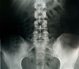
X-Ray
X-ray is often used for cancer diagnosis, staging and treatment. An X-ray uses electromagnetic radiation to make images. In low doses, X-rays can be used to construct images of structures inside the body to detect and stage a tumor. In higher doses X-rays can be used in radiation therapy to help destroy cancerous cells in the body.

Ultrasound
Ultrasound uses high-frequency sound waves to produce images of organs and tissues within the body. By capturing images in real time, ultrasound exams reveal the structure and movement of the body's internal organs. Ultrasound can also be used to precisely locate the position of a tumor in order to guide a biopsy or aspiration procedure. For example, ultrasound may be used to mark out the boundaries of a tumor prior to its removal. It can also be used to administer cancer treatments.Depending on the body part being examined, some types of ultrasound include: abdominal, breast, cardiac, pelvic, prostate, renal, testicular and thyroid. Unlike X-rays, ultrasound exams do not use radiation.

Positron Emission Tomography (PET)
Positron emission tomography (PET)is a nuclear medicine, functional imaging technique that produces a 3-D image of functional processes in the body. The system detects pairs of gamma rays emitted indirectly by apositron-emitting radionuclide (tracer), which is introduced into the body on a biologically active molecule. 3-D images of tracer concentration within the body are then constructed by computer analysis.

Computed Tomography Scan (CT Scan)
CT Scan is an imaging test that uses x-rays to create cross-sectional pictures of the internal organs of the body. Its is sometimes used with a safe inter venous dye called a “contrast,” which is injected to the body before the test. This contrast dye highlights specific areas inside the body, which gives an exact and clearer image if the internal organs and tumors if any.
How Does a CT Scan Diagnose Cancer?
You will be asked to lie down on a narrow table that slides into the center of the CT machine.
Once you are inside the scanner, the beam rotates around you as you lie still and relaxed.
A computer then takes this information and uses it to create many images, called slices. These images are stored and viewed on a computer monitor, or can be printed on film. The Computer can also take these slices and create a 3d view of your chest or abdomen.
The scan only takes only a few minutes. The newest multidetector scanners can image your entire body in less than 30 seconds.
How to Prepare for the Test
Your doctor will give you specific instructions on how to prepare for the Thoracic CT. Usually you will be asked not to eat or drink anything for 4-6 hours before the test.
If you have an allergy to IV your doctor can offer alternatives such as drinking the contrast dye or via an enema. Speak to your physician about what is the most effective way to do the test he will be happy to give help be as comfortable as possible.

Magnetic Resonance Imaging (MRI)
Magnetic resonance imaging (MRI) is a test that uses a magnetic field and pulses of radio wave energy to make pictures of organs and structures inside the body. In many cases MRI gives different information about structures in the body than can be seen with an X-ray, ultrasound, or computed tomography (CT) scan. MRI also may show problems that cannot be seen with other imaging methods.
For an MRI test, the area of the body being studied is placed inside a special machine that contains a strong magnet. Pictures from an MRI scan are digital images that can be saved and stored on a computer for more study. The images also can be reviewed remotely, such as in a clinic or an operating room. In some cases, contrast material may be used during the MRI scan to show certain structures more clearly.
See pictures of a standard MRI machine and an open MRI machine .
Why It Is Done?
Magnetic resonance imaging (MRI) is done for many reasons. It is used to find problems such as tumors, bleeding, injury, blood vessel diseases, or infection. MRI also may be done to provide more information about a problem seen on an X-ray, ultrasound scan, or CT scan. Contrast material may be used during MRI to show abnormal tissue more clearly.
An MRI scan can be done for the:
-
Head
-
Chest
-
Blood vessels
-
Abdomen and pelvis
-
Bones and joints
-
Spine